Whilst I am no longer posting this blog at weekly intervals this definitely does not mean that my laboratory experiences are over – far from it!! I recently spent time with Dr. Rocio Finol-Urdaneta exploring aspects of electrophysiology.
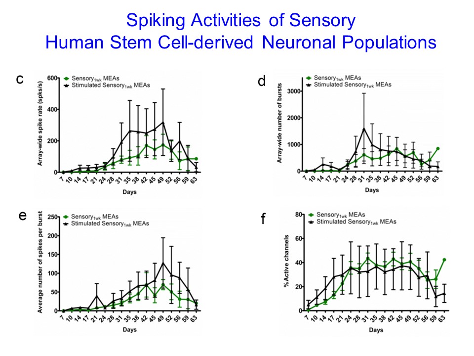
Looking down the microscope at the equipment set up to measure the spiking of the neurons in response to stimuli, I could see the pipette piercing a group of neurons. When the spiking occurred it was represented on the computer screen nearby – where the spiking levels and frequency were represented in graph form.
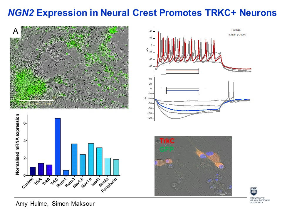
In the human body messages are passed by excitable tissue types, containing sodium channels e.g. muscle, neuron and secretory. The message spikes, which are measured by electrophysiology, are known as action potentials and represent a change in the electrical potential of the cell membrane.
When the cell is at rest the membrane potential of a living cell is negative. For example, when a muscle is relaxed it is said to be hyper-polarised or more negative.
As you can imagine, in such a complex biological structure as our body, the message transmission system is highly regulated. Body temperature plays an important part in this regulation process. At room temperature of @ 24 degrees the firing takes place approximately every 10 milliseconds, but at body temperature this firing level increases to every 1-2 milliseconds. To complicate matters further, in the spinal cord which is part of the nervous system, the spiking rate is slower than in the brain. Also, in the case of illness, changes in the message transmission rate can be observed.
So my time was well spent learning more detail about the role of electrophysiology in measuring the intricacies of the transmission of action potentials and the neurological characteristics that underlie them.