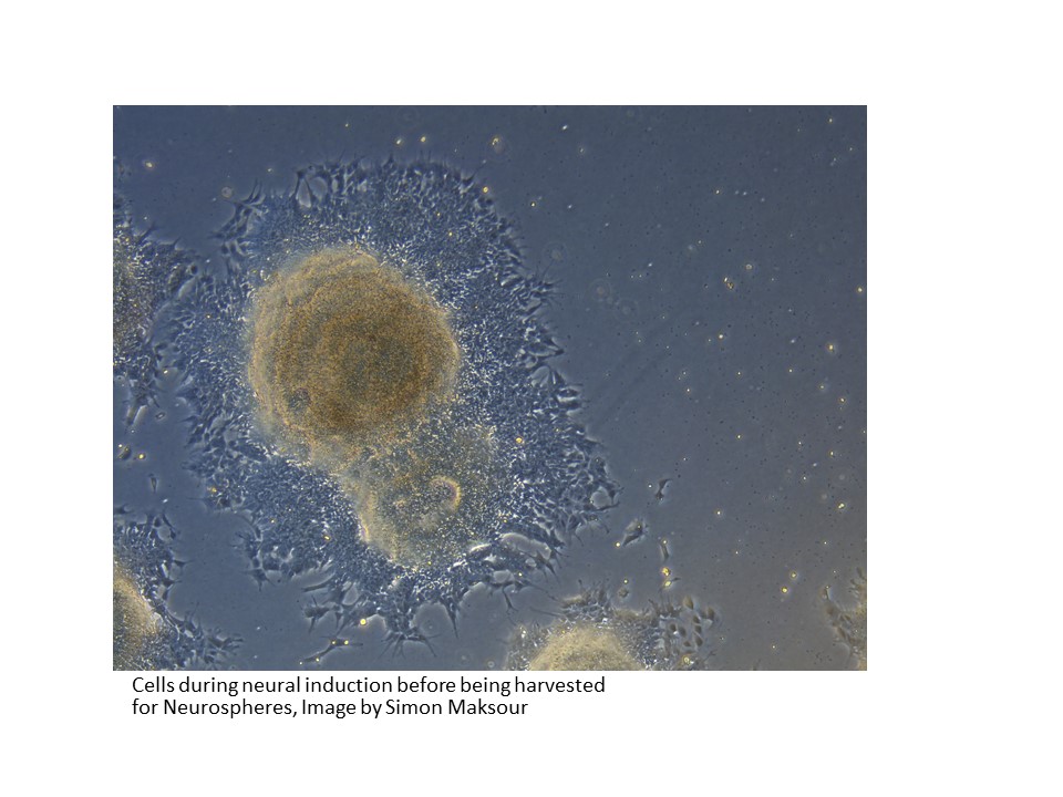
The Dottori laboratory not only cultures sensory neurons but also cortical neurons, that provide models for Alzheimers and Dementia research. The images below show 4 crucial stages in the process when culturing stem cells to generate cortical neurons:
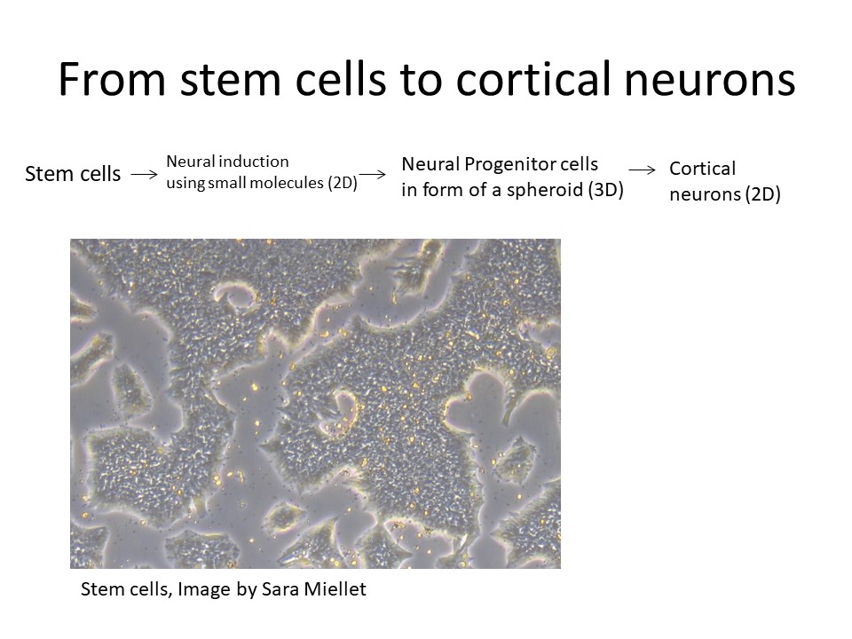
The first stage on the production of cortical neurons
The beginning of this process involves the isolation of pluripotent stem cells and their cultivation in the laboratory incubator.
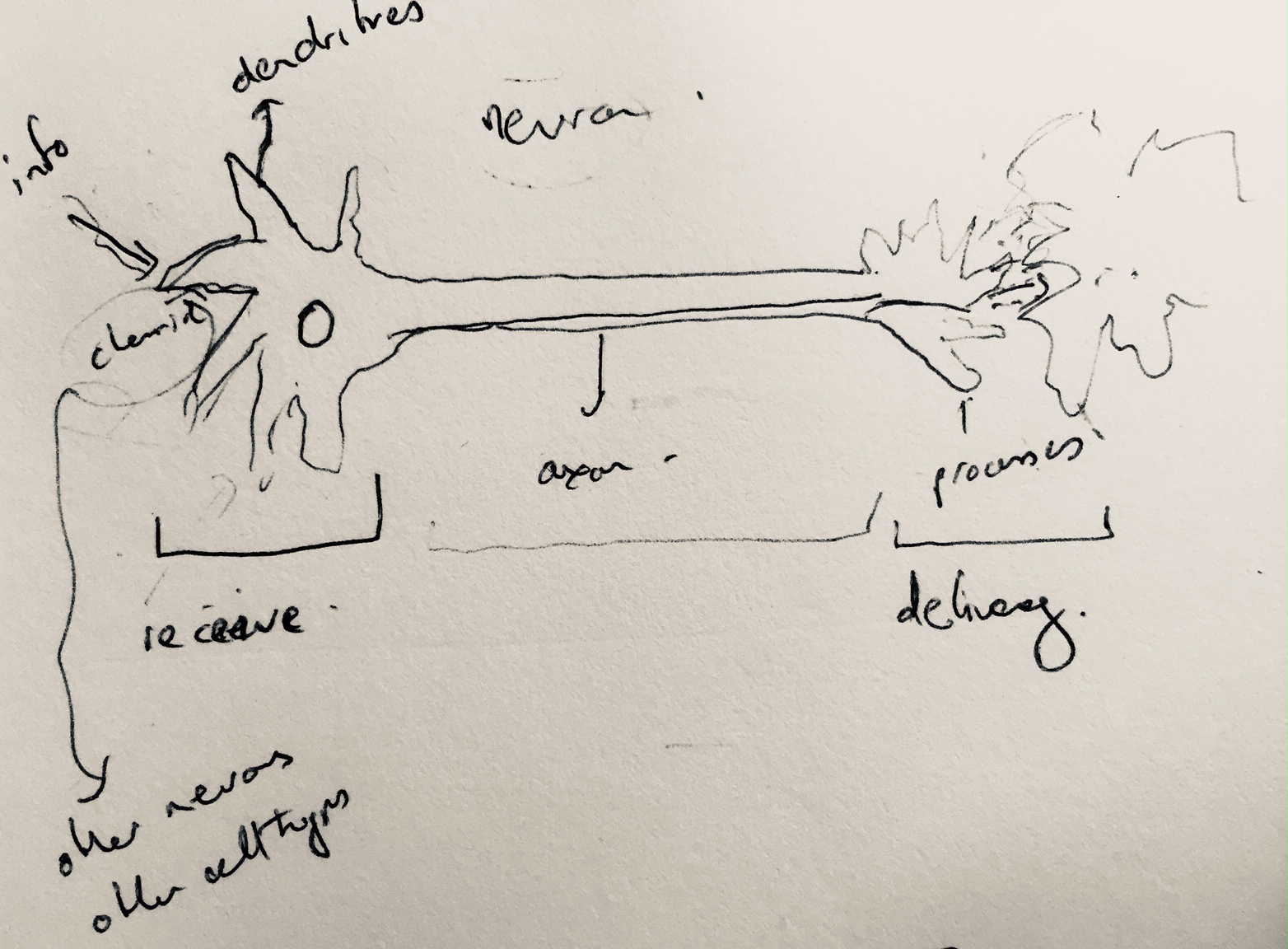
My distant memories of school biology lessons were revived when Mirella drew an impromptu diagram of basics of neuronal functioning in our body. This impromptu sketch helped her to explain to me that neurons communicate via chemical and electrical synapses, in a process known as synaptic transmission. They are thus categorised as electrically excitable cells housed in the human nervous system, whose function it is to process and transmit information.
Mirella’s spontaneous explanatory diagram of the structure of a neuron
As my laboratory observations and my discussions with Mirella continue my thoughts begin to turn to how I might recontextualise this complex scientific data to create interactive artworks in the future….??
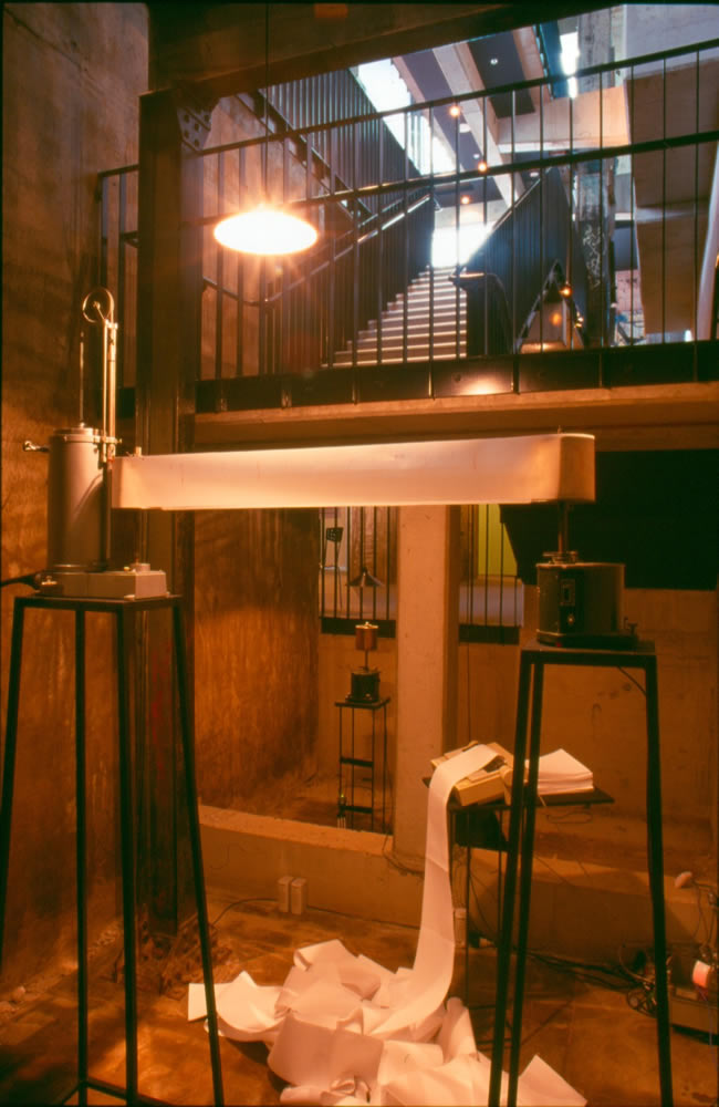
Temporal Intervals, 2003: A quasi-scientific installation I created @ the Brisbane Powerhouse exploring traces & remote Internet data transfer using antiquated scientific equipment (image, John Linkins)
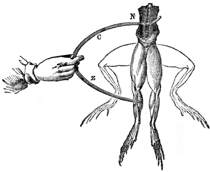
The neuronal responses remind me of my doctoral research project when I became interested in the so called ‘vital force’ possessed by the human body which could be regarded as an internal machine-like power known as ‘animal electricity’. I was particularly interested in the historical development of experiments that sought to identify and even locate this vital force.
This early research depended on the introduction of what were then cutting edge machines in the area of galvanics. Luigi Galvani (1737 – 1798) was an Italian physician, physicist, biologist and philosopher, who is credited with being the first to discover ‘animal electricity’ when he passed an electric current through frog’s legs, causing them to twitch. Initially known as ‘galvanism’, this is a forerunner of the contemporary scientific technique of electrophysiology .
Luigi Galvani – David Ames Wells, The science of common things: a familiar explanation of the first principles of physical science. For schools, families, and young students. Publisher Ivison, Phinney, Blakeman, 1859, 323 pages (page 290)
Subsequently, Carlo Matteucci (1811-1868) expanded on this research creating a ‘rheoscopic frog’, leading to the discovery, in approximately 1865, of a nerve’s action potentials by Julius Bernstein and Emil du Bois-Reymond. Currently, bio-electricity continues to be central to neurological experiments and is measured by techniques such as calcium imaging and electrophysiology.
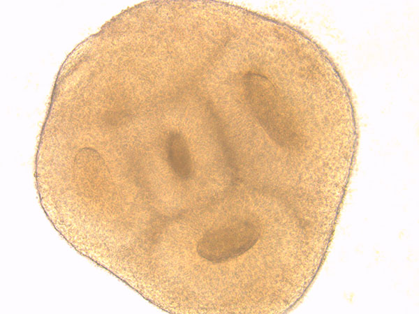
I was able to spend some time with Research Assistant: Sara Miellet, during which she explained more about the processes required to successfully culture organoids. As mentioned in my last post, organoids are small, three-dimensional structures derived from stem cells. They are useful to researchers because they mimic features of various selected organs in the human body.
Organoid developing, image Sara Miellet, Dottori lab
The Dottori laboratory is interested in modelling brain development by generating neural stem cells, derived from human stem cells, in vitro in the form of organoids for research purposes. The cells in an oganoid have been shown to have the remarkable ability to self-organise into complex structures.
The image above shows an example of cellular self-organisation in a developing neuronal organoid. These structures are called neural rosettes because they closely resemble the formation of the neural tube in a developing human embryo. This neural tube subsequently evolves in to the embryonic brain and spinal cord. Continue reading Continuations→