I was able to spend some time with Research Assistant: Sara Miellet, during which she explained more about the processes required to successfully culture organoids. As mentioned in my last post, organoids are small, three-dimensional structures derived from stem cells. They are useful to researchers because they mimic features of various selected organs in the human body.
The Dottori laboratory is interested in modelling brain development by generating neural stem cells, derived from human stem cells, in vitro in the form of organoids for research purposes. The cells in an oganoid have been shown to have the remarkable ability to self-organise into complex structures.
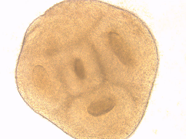
The image above shows an example of cellular self-organisation in a developing neuronal organoid. These structures are called neural rosettes because they closely resemble the formation of the neural tube in a developing human embryo. This neural tube subsequently evolves in to the embryonic brain and spinal cord.
Interestingly however, there is a limit to how big researchers are able to grow organoids in the dish. This is because they lack a vascular system to provide nutrients and oxygen to the centre of each organoid. Sara explained that, as the period of time in the incubator increases, the cells at the centre of the organoid are starved of these necessary nutrients and will die;
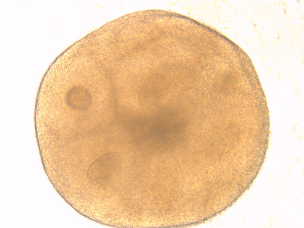
This can be seen beginning to take place in the image above in the form of a dark central group of cells. To prevent cell death and to grow larger organoids a specially designed incubator that agitates the cultures is used. Agitation helps the life-giving nutrients in the culture dish to reach the centre of the cultures, thus prolonging their life.