Returning to some earlier research in more detail I have been investigating neuronal firing.
As has been previously discussed in earlier posts, neuronal firing is the name given to the changes that take to enable neurons to communicate with each other. This neuronal communication occurs via electrical impulses and neurotransmitters.
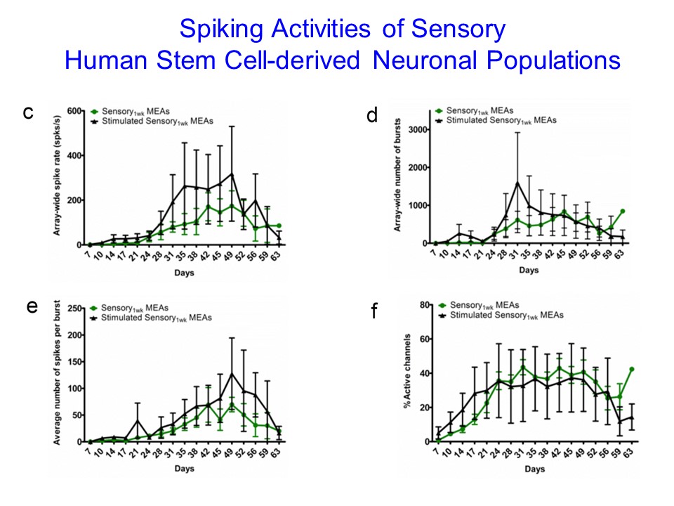
As you can see from the chart above, the activity of the cultured sensory neurons can be measured in a number of ways relating to the bursts of activity, spike rate and active channels.
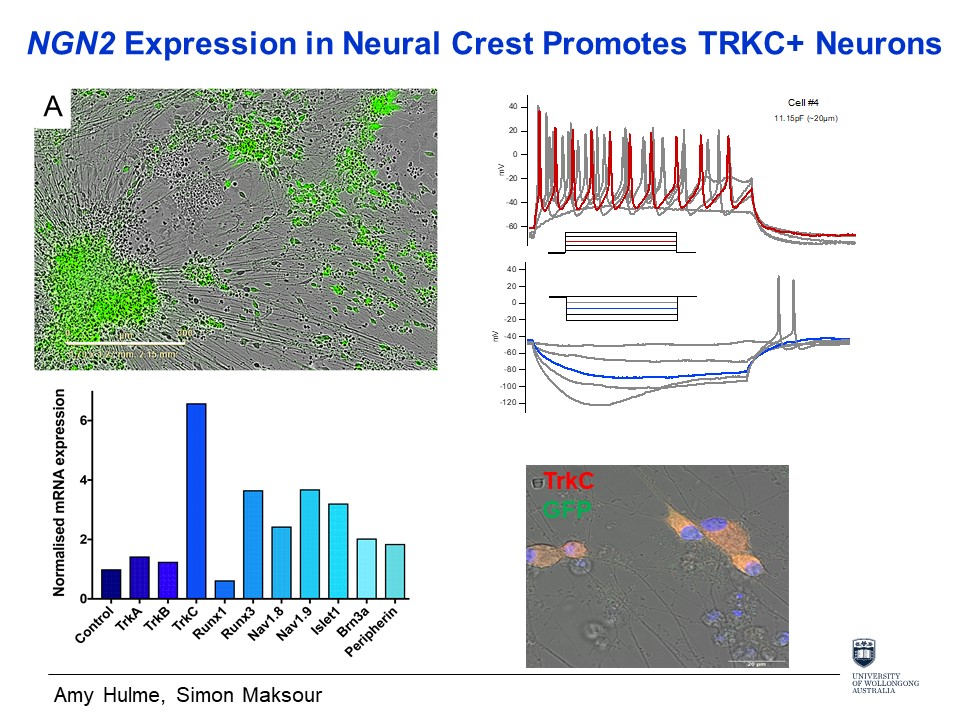
The diagram above represents the real-time firing of the cluster of neurons. The electrical impulse, known as an action potential, causes part of the neural membrane to open and allow positively charged ions into the cell whilst releasing negatively charged ions; causing a positive charge.
Using my own cells has been central to a number of my previous art/science collaborative projects i.e. my landmark use of cardiac cells cultured from my own adult pluripotent stem cells in 2002-3. After careful consideration, I decided not to follow this methodology for this Synapse residency – in part because the Dottori lab are already using human stem cells and, even in 2019, the ethical clearance for my own cells at UoW was proving difficult; and partly because this methodology seems to have become almost like a default process for artists in the intervening years since the ‘machina carnis’ project.
This trope is always at the back of my mind tho and recently I have begun to wonder how the responses of animal neurons would differ from the Dottori laboratory’s human sensory neuronal firing results? Apparently very little at this laboratory research phase. The difference between human and animal results becomes manifest in clinical trials, where a drug that has given promising results during animal testing proves disappointing when applied to humans.